
概要
临床表现。
NDP相关的视网膜病是以一系列视网膜血管和纤维渐进性病变为病理特征,表现为出生发病,程度进展性的视力受损。最严重的表型被称为“早产儿视网膜病”(ND), 病理特征是继发于视网膜血管发育不良和视网膜剥离的灰黄色纤维组织增生(假性神经胶质瘤)。先天性失明几乎出现于所有病例。大约30-50%的男性ND病人会伴有发育迟缓/智力障碍, 行为异常,或表现出精神病样症状。绝大多数的男性病人会有感音神经性耳聋。较轻的症状包括:原发性持续性玻璃体增生(PHPV), 表现为大量渗出物形成的从视盘到晶体的白色条状物; X-连锁 家族性渗出性玻璃体视网膜炎(XL-FEVR), 表现为视网膜周围血管异常伴或不伴纤维化改变和视网膜剥离;早产儿视网膜病(ROP)和渗出性视网膜病,一种渗出性、增生性的血管病变。家庭成员之间的表现型可能变化较大。
疾病管理。
对症治疗: 对于非完全性视网膜剥离,可以用手术和/或激光治疗,早期治疗有可能改善预后。手术对于眼压增高有帮助。为了控制疼痛而摘除眼球的情况很少见。听力的治疗可以用助听器和植入人工耳蜗。行为异常和认知障碍的问题需要对应的支持治疗和干预。
遗传咨询。
NDP相关的视网膜病呈X-连锁遗传。 男性病人不会将其致病性变异传给儿子,但会遗传给女儿,使其成为携带者。女性携带者有50%几率将其致病变异遗传给子女; 得到致病变异的儿子将是受累的患者,女儿将继续携带,一般不发病。如果家系的致病突变已经明确,女性亲属的携带者检测和孕妇的产前诊断是可行的。
GeneReview Scope
| NDP相关的视网膜病: 含表型 1 |
|---|
|
诊断
提示性发现
NDP相关的视网膜病患者应当有如下眼部特征:
- 先天性视觉丧失/失明
- 双侧, 通常是对称性的眼睛受累
- 原发性持续性玻璃体增生, 透明小管, 浅前房, 玻璃体视网膜出血。小眼球和白内障可能会出现
- 出生时表现晶体后纤维化和视网膜血管病变(白瞳症),儿童期直至青春期呈进展性改变
NDP 的致病变异导致的视网膜病变谱可以从典型的早产儿视网膜病 (ND) 到 X-连锁 家族性 渗出性玻璃体视网膜病 (FEVR), 以及部分表现为原发性持续性玻璃体增生 (PHPV), 渗出性视网膜病和进展性的早产儿视网膜病(ROP)。 各种表型可能为连续的视网膜改变并有较多的交叉(表 1)。
表 1。
NDP的致病变异相关的眼部表型分类
| 表型 | 眼部表现 / 发病年龄 | 进展 / 年龄 | 视觉 |
|---|---|---|---|
| 早产儿视网膜病 (ND) | 晶体后浅灰色-黄色纤维血管团块 ("假性神经胶质瘤") , 视网膜剥离 (常见) / 出生至 3 月 | 白内障, 眼后部粘连 (虹膜到晶体), 眼前部粘连 (虹膜到角膜), 虹膜萎缩, 前房变浅, 角膜浑浊, 带状角膜病, 眼压低, 眼球萎缩 (眼球痨) / 3 月 到 8-10 岁 | 光感受损或缺失 |
| 原发性持续性玻璃体增生 (PHPV) 1 | 纤维化白色条索伴随透明小管从视盘延伸至晶体后囊 / 出生 | 未知 / 未知 | 不同程度受损 |
| X-连锁 家族性 渗出性玻璃体视网膜 (FEVR) 2 | 视网膜外周暂时性无血管区 ± 先天的 视网膜折叠, 黄斑异位, 锯状缘纤维组织带 / 出生时 | ± 视网膜剥离 (牵拉性 和/或 渗出性; 可能单侧) / ≤ 20 岁 | 正常或受损 |
| 早产儿视网膜病 (ROP) 4B/5期 3 | 视网膜新生血管纤维性增殖, 末期晶体后纤维素增生 / 早产儿出生时 | 部分或完全性视网膜剥离 | 受损或失明 |
| 渗出性视网膜病 4 | 单侧视网膜毛细血管扩张, 纤维性渗出 | 进行性血管性渗出, 视网膜下渗出、纤维化, 视网膜剥离 | 正常或受损 |
PHPV中的NDP基因致病变异 [Shastry 2009a]
2.
NDP基因的致病变异在临床诊断为X-连锁家族性渗出性玻璃体视网膜病FEVR中并不罕见[Nikopoulos et al 2010a, Nikopoulos et al 2010b, Poulter et al 2010, Walsh et al 2011, Yang et al 2012].
3.
表现为ROP表型的病患中NDP的致病变异很罕见[Shastry et al 1997]。 后续研究发现NDP基因 编码区外的变异很可能是多态和/或者NDP基因表达的调控因素[Kenyon & Craig 1999, Hiraoka et al 2001a, Talks et al 2001, Haider et al 2002, Hutcheson et al 2005, Shastry 2009b]。
4.
在携带有NDP致病性变异的家系中,女性携带者可以有镶嵌体的表型(渗出性视网膜病); 她的儿子则会表现为典型的早产儿视网膜病[Black et al 1999]。
确诊
NDP相关的视网膜病的诊断需要在 先证者 找到NDP基因的 致病性变异 (详见 表2)。
连锁分析。在非常罕见的疾病家系中若未发现NDP致病性变异, 应当考虑用连锁分析对多于一个受累的患者家系进行检测。 连锁分析需基于已对受累家系成员完成了确定的NDP-相关视网膜病的临床诊断并且对家系个体间的遗传学关系很清楚的前提下进行的。连锁分析的结果依赖于待测家系成员的同意和配合。用于NDP 连锁分析的标记紧密连锁与NDP位点可以提供充分的信息;因此,可以用于很多家系的NDP相关的视网膜病的连锁分析,准确率高于95%。在能够提供足够信息的家系, 连锁分析可以确定可疑女性携带者的携带状态.
表 2.
NDP相关的视网膜病分子遗传学检测小结
2.
序列分析可以检测良性, 可能良性, 意义不确定, 可能致病, 或致病变异. 致病突变可能包括基因内缺失/插入和错义, 无义, 和 剪接位点 变异; 但是, 外显子 或全基因缺失/重复是不能被检测的. 对于序列分析报告结果的解释的相关问题, 详见此处。
3.
4.
包含亚显微水平的缺失致病突变,涉及部分或全部NDP基因和临近的基因组的序列占所有致病突变的15%[Berger & Ropers 2001; Sims,未发表]。
5.
6.
检测外显子或全-基因缺失/重复,这些变异是不能用仅检测编码区和基因组的DNA上的内含子的(侧翼区)的序列分析方法进行的。在众多的技术手段中: 定量PCR, 长片段 PCR, 多重连接依赖的探针扩增反应(MLPA), 及预设特定染色体区域探针的染色体芯片(CMA)可以用于此部分变异的检测。
7.
亚显微水平的缺失包含部分或全部NDP基因和邻近基因组的区域[Berger & Ropers 2001; Sims, 未发表]。
临床特征
临床描述
带有NDP基因的致病性变异 的男性患者的眼部表现通常为双侧对称性的。出生即发病,呈进展性。典型的早产儿视网膜病 ND 表型 是在该病被命名后首先被描述的也是最为详尽刻画的眼部表现. 随着NDP基因的发现和分子遗传学检测的临床应用,很显然NDP相关的视网膜病包括早产儿视网膜病(ND),原发性持续性玻璃体增生 (PHPV), 和 X-连锁 家族性家族性渗出性玻璃体视网膜病 (XL-FEVR), [Riveiro-Alvarez et al 2005, Dickinson et al 2006, Kondo et al 2007] (表1)。
眼部表型在同一个家庭中可能也有差异[Berger & Ropers 2001, Allen et al 2006]。已报道一个家系中有9个受累的男性患者,不同人眼部表现有3-4岁单侧次视网膜剥离缓慢发展为快8岁时的牵拉剥离,也有男性到79岁时才表现为视网膜周围色素改变[Allen et al 2006]。
新生儿视网膜病
- 眼部发现。 出生时眼球, 虹膜、前房、角膜、眼压和眼球体积可正常,新生儿和婴儿期, 典型表型是视网膜灰黄色、反光、凸起的团块, 透过清晰的晶体可以发现。这些团块因为类似肿瘤被称为 "假性神经胶质瘤"。部分或全部视网膜剥离出现在几个月内。
从婴儿期到儿童期,典型的进展症状包含: 晶体混浊(白内障), 虹膜萎缩,粘连出现在晶体和虹膜间 (后房粘连) 和虹膜与角膜间 (前房粘连),前房变窄,流出道堵塞导致眼压增加和疼痛。
角膜浑浊、带状角膜、眼压缺失和眼球萎缩 (眼球痨)是后期症状。在早产儿视网膜病进展末期,视网膜变为乳白色不透明,眼球变小沉入眼眶[Drenser et al 2007]。 - 认知/行为问题。约 30%-50% 的早产儿视网膜病ND男性患者的表型有发育迟缓/智力障碍,可能出现轻微的行为异常或者精神症状。家系内和家系间差异和行为问题的表现与否及程度差异普遍存在。严重的神经病学表型如婴幼儿痉挛已有报道[Lev et al 2007]。
- 听力问题。绝大多数早产儿视网膜病ND男性患者的表型 会在婴幼儿时期出现进展性的感音神经性耳聋(表 1)。耳聋可以表现为隐匿起病。
听力学测试病变出现在耳蜗(特别是血管纹),蜗后和脑感音系统功能正常。早期的耳聋是轻度、不对称性感音性聋,高频听力损失出现在青春期。到35岁可表现为重度、对称性的宽波段耳聋。语言功能可以相对较好的保留下来,即使阈值损失比较严重。[Halpin et al 2005, Halpin & Sims 2008]。
大多数受累的患者对先天的失明的适应比对迟发进展性的耳聋更容易。
- 其他.周围血管病 (图 2)在较多男性受累的患者中可能是相关的临床表现[Smith et al 2012]。
肺动脉高压曾被报道过。
整体的健康状况正常。寿命可能由于疾病症状带来的风险而受影响,如智障、失明和/或耳聋等增加了受伤、吸入性肺炎和癫痫并发症。
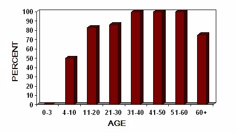
图 1.
早产儿视网膜病男性患者听力受损情况。一组 (56例) 男性病人耳聋情况的年龄分组 [Smith et al 2012]
图 2.
早产儿视网膜病男性患者外周血管病。一组 (56例) 男性病人耳聋情况的年龄分组 [Smith et al 2012]
原发性持续性玻璃体PHPV的特征是伴随血管的纤维白色条索从视盘延伸至颞后晶状体囊。视网膜折叠或剥离;晶体可能变混。尽管完全性视网膜剥离有过报道,但是并不是所有病例都会进展至此[Wu et al 2007]。原发性持续性玻璃体PHPV通常表现为单侧; 因此双侧病变提示NDP相关的视网膜病变。
X-连锁家族性渗出性玻璃体FEVR 的特征是视网膜血管形成的过早停止,从而导致视网膜周边出现无血管区。无血管区可以作为单独症状也可以伴随先天的视网膜镰刀状折叠或剥离。当出现视网膜镰刀状折叠时,黄斑可能会被牵拉移位(此为黄斑异位)。
这些眼部表现可能会进展为视网膜剥离,一方面的原因是来自颞侧视网膜周边进展性纤维化而增加的牵引力,另一方面原因是异常的视网膜外周血管形成而出现的脆性毛细血管的渗出。视网膜剥离通常由于黄斑受累而伴随出现中心视力。
进展性早产儿视网膜病(ROP;4B/5期) 视网膜改变类似于家族性渗出性玻璃体视网膜病FEVR。曾在16个有进展性早产儿视网膜病ROP的早产儿当中,4个被检出NDP基因致病突变[Hiraoka et al 2001a], 提示是否NDP 致病突变可能在早产儿中更容易导致早产儿视网膜病ND眼部表型。近年在17个进展性早产儿视网膜病ROP患儿中, 只有1个被检出NDP致病性变异,位于5’UTR区[Hiraoka et al 2010]。在科威特102例早产儿的研究中,仅有多态位点被检出,并未发现致病突变[Haider et al 2001]。 Hutcheson et al [2005]Hutcheson等研究了来自不同种族的严重早产儿视网膜病ROP (≥3期),仅在NDP基因的非编码区(UTR)发现5例变异。目前尚无这些NDP基因多态位点在早产儿视网膜病ROP中的致病证据。
渗出性视网膜病是一种渗出行的血管增生病变,20岁前发病,常见婴儿和儿童病患。男女病患比约为10:1。视网膜病变包括毛细血管扩张,静脉和毛细血管呈纺锤形扩张,微动脉瘤。视网膜下渗出和出血可见到,常位于黄斑和/或颞上区域。渗出性视网膜剥离和视网膜毛细血管灌注减少可能出现。其他并发症包括虹膜睫状体炎,白内障和新生血管青光眼。超过90%的报道病例为单侧受累。
组织病理学
视网膜血管病可能是原发性的病理生理改变,导致了随后的纤维化改变和玻璃体出血。视网膜神经节细胞缺失也可能发生。异常的视网膜血管形成已在小鼠模型中被报道[Richter et al 1998, Schäfer et al 2009]。在早产儿视网膜病的小鼠模型中, Rehm et al [2002]报道了进展性的位于耳蜗血管纹的血管缺失,导致听力受损。在Ndp敲除小鼠中,基因 表达的差异研究发现了视网膜(光感受器) 表型的致病基因[Lenzner et al 2002]。小脑血管改变在Ndph敲除鼠中也被报道过[Luhmann et al 2008]。
杂合突变
在极个别情况下,女性携带者可能有视网膜病变(视网膜剥离,异常的视网膜血管形成) [Lin et al 2010, Parzefall et al 2014],相关的视力受损也被报道过[Yamada et al 2001]。一个严重的眼部表型在一个家系中的多名女性中曾被报道过[Khan et al 2008]。
一些女性携带者可能表现出轻度的感音神经性耳聋[Halpin et al 2005; Sims, 未发表]。疾病表型曾在两个X染色体与常染色体易位的女性中被报道 [Meire et al 1998]; 然而, 携带者 出现表型很罕见,通常被假设归因于X-染色体非随机失活。
基因型-表型相关性
家系内和家系间病例的表型差异已被发现,不管是视网膜表型 [Wu et al 2007],还是非视网膜表型[Khan et al 2004, Allen et al 2006]。自从在两兄弟带有启动密码子的NDP基因缺失中发现智力障碍表现不同后被引起注意[Zhang et al 2013], 尽管其他报道的较多兄弟案例中典型的NDP视网膜病变均表现出智障[Arai et al 2014]。更多其他表型包括小脑萎缩,运动障碍和智障在三对兄弟中的报道也支持其他非NDP致病性变异的表观遗传学因素可能影响临床表型的假说[Liu et al 2010]。
男性患者若有基因缺失,会表现出比非缺失突变病人更加严重的表型[Suárez-Merino et al 2001, Wu et al 2007, Yang et al 2012, Arai et al 2014]。 对于NDP基因缺失型早产儿视网膜病ND患者,除了眼部表现外,可能出现小头畸形、重度到极重度智力障碍、癫痫、肌阵挛、体细胞生长障碍和/或青春期延迟。破坏了半胱氨酸结模序的致病突变可能跟严重的视网膜发育不全相关[Wu et al 2007]。在四个严重的视网膜发育不良的病人中发现的独特的单核苷酸变异改变了半胱氨酸残基,可能改变了蛋白质的重要结构[Drenser et al 2007]。尽管部分案例提示
错义变异导致蛋白质C端氨基酸改变可能与轻型家族性渗出性玻璃体视网膜病 FEVR表型相关[Allen et al 2006], 但是也有大量的数据显示存在严重眼部表型,伴或不伴智力障碍的病例检出导致蛋白质C端氨基酸改变的错义突变[Sims, 未发表]。
目前并无证据支持单碱基致病突变和认知障碍或者听力受损相关,有一个病例检出单碱基突变(c.134T>A),被报道了严重的神经疾病表型,包括极重度智障和婴儿肌阵挛[Lev et al 2007]。据报道有较多并且差异明显表型邻接
基因综合征往往可见NDP+缺失[Sims et al 1989, Rodriguez-Revenga et al 2007]。有报道显示一个家族性特发性肺动脉高压家系中存在X染色体上p11.3-11.4的微缺失,包含NDP基因[Staropoli et al 2010]。
有一个案例显示, 受累的儿子有复杂的表型(脑与视网膜微血管病伴随钙化和囊肿),母亲被诊为单侧不典型渗出性视网膜病,有NDP基因的致病错义变异和CTCI基因的复合杂合变异[Romaniello et al 2013]。CTCI基因已知与脑脊髓微血管病伴钙化和囊肿综合征[Anderson et al 2012]和脑视网膜微血管病伴钙化中的微血管病变相关[Polvi et al 2012]。这一报道提示两个基因之间可能存在相互作用,从而导致携带者母亲和先证者均出现严重和复杂的表型。
术语
早产儿视网膜病有如下曾用名:
- 遗传学眼球萎缩
- 假性神经胶质瘤
- 埃皮斯科皮失明
患病率
目前尚无患病率或发病率数据。一项对多样性儿童玻璃体视网膜病的研究显示NDP基因的致病突变率11/109[Wu et al 2007]。早产儿视网膜病在不同种群中均有报道
, 如北欧和中欧、欧裔美洲人、非裔美洲人、法裔加拿大人、西班牙人、中国人和日本人。尽管并没有明显高发的族群,但是在早产儿视网膜病被首次描述后的十年内报道的病例大多数来自于斯堪的纳维亚。遗传性 (等位基因) 疾病
除了在此GeneReview 词条提及的表型外,没有其他已知症状与NDP基因突变相关;尽管曾在一个X-连锁青少年视网膜劈裂的家系中发现,NDP致病性变异 与 RS基因致病突变共分离[Hiraoka et al 2001b],但是在其他的家族性或散发性视网膜劈裂病例中并未发现[Shastry et al 2000]。
鉴别诊断
在早产儿视网膜病(ND)先证者中,如果眼部的主要症状是单侧假性神经胶质瘤,视网膜母细胞瘤(RB)常作为疑似诊断。因为早产儿视网膜病ND常为双侧发病,视网膜母细胞瘤RB的诊断并不需要经常考虑。有经验的眼科医师可以通过眼底检查对两种疾病进行鉴别。
早产儿视网膜病变ND可能与原发性持续性玻璃体增生PHPV和早产儿视网膜病 (ROP)类似 (尤其是经过辅助给氧治疗的早产低体重儿)[Shastry 2009a]。
常染色体显性家族性渗出性玻璃体(adFEVR)是另一种需要鉴别诊断的疾病。NDP基因编码卷曲蛋白-4 (Fzd-4) 的配体,活化经典的Wnt信号通路 [Xu et al 2004, Hendrickx & Leyns 2008]。 FZD4 [Robitaille et al 2002]、 LRP5 [Jiao et al 2004, Toomes et al 2004]和 TSPAN12 [Nikopoulos et al 2010b, Poulter et al 2010, Yang et al 2011] 基因的致病变异已被证实与常染色体显性家族性渗出性玻璃体视网膜病FEVR相关。由于FEVR的部分表型与早产儿视网膜病ND交叉, FZD4、LRP5和TSPAN12 基因的致病变异可能与某些遗传模式不是很明确的早产儿视网膜病病例相关。
伴随原发性持续性玻璃体增生症PHPV的视网膜发育不良可能与Walker-Warburg syndrome肌营养-肌营养不良蛋白聚糖病的无脑回畸形相关, 后者是一种常染色体隐性遗传形式的先天的肌营养不良,常伴随13号染色体三体的多种畸形出现。这些都不会给早产儿视网膜病的诊断造成干扰。对于没有眼部表现的智力障碍/或进展性感音神经性耳聋 (详见耳聋综述),早产儿视网膜病的诊断一般不予考虑。
疾病管理
初诊后评估
为了确定疾病的严重程度和所需的处理方式,诊断为NDP相关的视网膜病的患者, 需要进行如下评估:
- 完整的眼科检查
- 基本的听力学评估
- 如果儿童早期发育标志未明确的话,需要进行神经发育评估
- 必要时进行行为评估
- 临床遗传咨询
对症治疗
眼部症状
- 大部分的早产儿视网膜病 (ND)男性患者出生即有完全性视网膜剥离的表型,对于保护视力的干预治疗并无太大帮助。眼科检查是有必要的。
- 没有完全性视网膜玻璃的患儿,早期的手术或激光治疗对于改善预后或许有益。
- 曾有报道Chow et al [2010] 早产儿视网膜病男婴(孕37周出生),经过成功激光焊接治疗后,23月龄时Teller 视敏检测右眼视力0.2
- 在一份回顾性病例综述研究中,接受了早期玻璃体切除术的14位早产儿视网膜病患儿的三级护理病例 (1988-2008年) 显示有一半 (7/14 病例) 的患儿在12月龄 (平均 4.5 月龄) [Walsh et al 2010]。
- 在早产儿视网膜病的表型的进展期,眼压增高可能需要手术。因缓减疼痛而进行眼球摘除的情况罕见。
感音神经性耳聋
- 助听器对于儿童及成人有较大帮助。
- 人工耳蜗必要时可以考虑。
行为异常是许多早产儿视网膜病患者及其监护人的终生问题,不管智障及认知异常有无出现。干预和治疗对于支持和提供最大限度的受教育机会有帮助。尽管并无研究支持特定药物对于治疗早产儿视网膜病有益,经验显示精神类药物或许有所帮助。
生存状态监管
以下措施当被建议:
- 即便视力已经严重下降,所有的NDP相关的视网膜病患者仍应进行常规的眼科随访。
- 若NDP相关的视网膜病患者已经失明,常规的的听力检查可以早期发现并适当干预听力问题。
- 临床观察当注意淤血和溃疡的发生
应当避免的事件/环境
因为耳聋风险较高,应当避免接触严重噪音。
妊娠管理
曾报道一例携带有NDP致病性变异的男性胎儿在妊娠34周时被娩出(妊娠28周及33周时超声检测排除视网膜剥离)[Sisk et al 2014]。生后2天接受双眼无血管视网膜激光消融治疗,10月龄随访双眼视网膜仍完全附着[Sisk et al 2014]。
治疗及疗效
有报道显示在视网膜血管生成因子被敲除的小鼠中通过晶体转入norrin蛋白编码基因而纠正表型 Ohlmann et al [2005]。作者也提及了norrin蛋白对视网膜神经节细胞的增殖存在潜在刺激作用。对于疾病的
临床研究点击 ClinicalTrials.gov。
遗传咨询
遗传咨询是为个人或家庭提供遗传病自然病程、遗传规律及专业建议,从而使其知情后做出医学和个人决定的过程。以下内容将从风险评估、家族史和家族成员遗传学检测等方面展开。该内容不一定适合所有需要面对或解决的个人、文化或者伦理问题时的遗传学专业咨询。—ED.
家族成员风险
男性 先证者 父母
男性 先证者 的后代。带有NDP相关的视网膜病致病变异的男性会将该 NDP致病性变异 遗传给女儿, 使其成为携带者, 而儿子不会遗传。
男性 先证者 的其他家族亲属。先证者的姨可能是 NDP致病性变异 的携带者,他们的子女可能根据性别的不同成为携带者或 受累的 的患者。
携带点检测
确定女性携带者应注意:
- 首先家族中要确定 NDP致病性变异 ;
或者 - 以下情况进行连锁分析:
- 家族成员规模适合进行连锁分析;
并且 - 家族成员可以进行配合检测。
注意: X-连锁 遗传病的携带者 是杂合突变,很少出现相关临床表现; 但是也有部分病例被报道过 [Lin et al 2010, Romaniello et al 2013, Parzefall et al 2014] 。因此,当在遗传咨询或产前咨询中讨论 携带者 风险时,此类情况应被提及。
相关的遗传咨询问题
家族计划
DNA库 是储存 DNA (常用白细胞提取) 已被将来所用。 因为检测方法和我们对基因的认识、等位基因变异以及疾病的认识可能会随着时间发生改变,对于 受累的 个体,应当考虑储存 DNA。
相关资源
GeneReviews 团队选择以下特定疾病组织和/保护伞组织和/或患者及其家属的疾病注册机构。GeneReviews 不对其他组织提供的信息负责。对于信息的入选标准, 请点击此处.
- National Library of Medicine Genetics Home Reference
- Norrie Disease Association (NDA)PO Box 3244Munster IN 46321Email: joinnda@gmail.com
- American Council of the Blind (ACB)2200 Wilson BoulevardSuite 650Arlington VA 22201Phone: 800-424-8666 (toll-free); 202-467-5081Fax: 202-467-5085Email: info@acb.org
- American Society for Deaf Children (ASDC)800 Florida Avenue NortheastSuite 2047Washington DC 20002-3695Phone: 800-942-2732 (Toll-free Parent Hotline); 866-895-4206 (toll free voice/TTY)Fax: 410-795-0965Email: info@deafchildren.org; asdc@deafchildren.org
- National Association of the Deaf (NAD)8630 Fenton StreetSuite 820Silver Spring MD 20910Phone: 301-587-1788; 301-587-1789 (TTY)Fax: 301-587-1791Email: nad.info@nad.org
- National Federation of the Blind (NFB)200 East Wells Street(at Jernigan Place)Baltimore MD 21230Phone: 410-659-9314Fax: 410-685-5653Email: pmaurer@nfb.org
- Norrie Disease RegistryMassachusetts General Hospital185 Cambridge StreetCRP Building North, 5th Floor, Suite 5300Boston MA 02114Phone: 617-726-5718Fax: 617-724-9620Email: ksims@mgh.harvard.edu
疾病分子遗传学
此处的信息和 OMIM 表格可能与 GeneReview其他地方不同: 会包含更多信息。-ED.
表 A.
NDP相关的视网膜病: 基因和数据库
表 B。
NDP相关的视网膜病的OMIM收录情况 (点击此处查阅所有信息)
分子遗传的发病机理
小鼠模型研究显示norrin蛋白缺乏在视网膜和耳蜗血管病变中至关重要[Xu et al 2004, Hendrickx & Leyns 2008],有可能也是导致人类早产儿视网膜病患者视觉受损及听力受损和/或智障等症状的血管生成的病理学原因。基因敲除小鼠(Ndp y/-) 表现出视网膜血管生成障碍,完全缺失视网膜毛细血管层,进展性的缺失耳蜗血管纹[Rehm et al 2002]。这些视网膜血管形成异常可能导致视网膜缺氧以及不可逆的视网膜损伤[Luhmann et al 2005]。
Norrin蛋白与Fzd4 CRD结合[Smallwood et al 2007]。 Norrin-Fzd4复合体启动信号通路[Ye et al 2010], 随后激活经典的 Wnt信号通路, 后者在视网膜血管发育过程中起着核心作用[Xu et al 2004, Warden et al 2007, Parmalee & Kitajewski 2008, Ohlmann & Tamm 2012]。参与内皮细胞增殖、迁移和视网膜浸润,参与动静脉分布,通过转录因子、细胞表面受体和细胞连接蛋白调控维持血脑屏障和血视网膜屏障稳定性[Paes et al 2011, Wang et al 2012]。 Norrin蛋白也通过激活Wnt/beta-catenin信号通路[Seitz et al 2010]和诱导Muller细胞神经生长因子合成[Ohlmann & Tamm 2012]而具有重要的神经保护作用。Norrin蛋白在星形胶质细胞和小脑贝格曼神经胶质细胞中广泛表达[Ye et al 2011], 提示可能在至今机制不明的中枢神经系统症状中起作用。
Norrin蛋白在智力障碍的发生过程中的作用仍然不清。近来, NDP 表达在视网膜前体细胞中因 Hedgehog (Hh) 介导 Gli2 结合于NDP基因启动子而开始[McNeill et al 2013]。作者考虑到除了Ndp在视网膜血管生成过程中的作用外,在神经前体细胞中的作用也被证明,因此提出假设:Ndp可能参与调控了更广泛的神经功能。
基因结构。NDP基因组的总长28 kb。cDNA包含3个外显子,编码区从第二个外显子的后半部开始,到第三外显子的前半部分结束,mRNA 全长 2.1 kb。第1外显子不编码,可能作为转录启动子区而使基因转录。早产儿视网膜病ND相关的致病突变可以出现在第一外显子[Schuback et al 1995, Kenyon & Craig 1999], 尽管功能仍有争议。一个被认为对蛋白质二级结构的维持起关键作用的多半胱氨酸区域存在于第三外显子编码的碳端。该区域正是大多数致病突变出现的地方,尽管其他编码区也都有散在分布。有关基因和蛋白的详细信息,详见 表A。良性变异。单核苷酸变异(SNVs) 引起或参与调控早产儿视网膜病ND相关症状的可能性仍然存在。位于第一外显子的插入 和 缺失 变异 与ROP相关性已被报道[Hiraoka et al 2001a, Dickinson et al 2006, Wu et al 2007]。由于第一外显子区域的插入和缺失也在较多的对照个体中出现[Sims,未发表], 提示可能属于良性变异。然而,这些位于第一外显子的变异仍然有可能与特定高危人群的临床血管病表型相关。
致病变异。已报道超过 100种NDP基因致病变异(错义, 无效, 剪切, 缺失)[Ye et al 2010]。大多数的致病突变是位于 NDP编码区的单碱基改变[Schuback et al 1995, Shastry 1998, Zaremba et al 1998, Black et al 1999, Hiraoka et al 2001b, Yamada et al 2001, Dickinson et al 2006, Drenser et al 2007, Kondo et al 2007, Lev et al 2007, Wu et al 2007, Khan et al 2008, Yang et al 2012]。
超过20种 DNA 重组也被报道过,包括基因内(小) 缺失, 或 亚显微("片段大于NDP基因") 缺失,大约占所有致病突变的15%[Berger & Ropers 2001; Rodriguez-Revenga et al 2007; Staropoli et al 2010; Sims, 未发表]。插入[Hiraoka et al 2001a],复杂重排[Schuback et al 1995], 以及X染色体易位[Meire et al 1998] 病例也均见诸报道。这些案例中的一部分被确认缺失范围超过NDP位点。这些病例的表型复杂,呈邻接基因综合征[Sims et al 1989, Suárez-Merino et al 2001, Rodriguez-Revenga et al 2007, Staropoli et al 2010]。曾有报道
Ke et al [2013] 依据蛋白晶体结构将NDP致病性变异分为5类: (1) 半胱氨酸残基, (2) 二聚体结合面, (3) 疏水核心, (4) Fzd4 结合位点,和(5) Lrp5/6 结合位点。 Fzd4 富含半胱氨酸结构域 (CRD) 的变异已被证明可以影响Norrin结合和信号转导[Zhang et al 2011]。
大多数的致病突变是罕见的,仅在单个病人/或家庭中被发现;少数致病突变在多个无关家系中出现, 致病错义变异均可能导致重要的氨基酸改变,常改变一连串半胱氨酸中的一个。这些半胱氨酸被认为与维持蛋白结构有重要作用,因此这些位置的致病突变可能对蛋白功能具有潜在的破坏作用。
正常基因产物。Norrin蛋白 [NM_000266; NP_000257] 是包含133个氨基酸的分泌蛋白;有半胱氨酸结模序(在许多生长因子中高度保守的结构)。晶体结构分析显示norrin蛋白以同源二聚体形式存在,后者是Frizzled-4 (Fzd4) 结合形式[Ke et al 2013]。复合体可以激活经典的Wnt/beta-catenin 信号通路,活化过程被Tspan12加强 [Junge et al 2009]。 Norrin蛋白对于中枢神经系统血管发育非常关键因为其在眼、耳和脑的血管生成以及维持血脑屏障(BBB)、血视网膜屏障(BRB)的稳定性维持中非常必要;且对视网膜神经元有神经保护功能。通过鼠、兔和人的原位杂交证实信使RNA存在于早期视网膜外颗粒层、内颗粒层和神经节层,提示其在视网膜发育过程中发挥作用
[Hartzer et al 1999]。后续深入研究 (参见 异常基因产物)也支持norrin蛋白在细胞或组织分化和维持细胞表型以及细胞间联系中发挥作用,对于正常的视网膜、中枢神经系统以及耳蜗的发育至关重要。
异常基因产物。Norrin蛋白缺陷小鼠模型(Ndp y/-) 的表型表现出视网膜血管发育受影响,视网膜表面毛细血管生长不足伴随视网膜内部毛细血管缺失[Richter et al 1998, Rehm et al 2002, Luhmann et al 2005, Ohlmann et al 2005] 和异常的玻璃体血管。迟发性耳聋会伴随耳蜗退化[Berger 1998]和内耳异常血管出现[Rehm et al 2002, Ohlmann et al 2005]。Ndp 敲除小鼠模型为视网膜血管发育中norrin蛋白的作用提供了更进一步的证据[Schäfer et al 2009, Ye et al 2011, Lee et al 2013]。外源性的norrin蛋白可以修复ROP小鼠模型的视网膜血管网
[Ohlmann et al 2005],减少血管缺失和病理性新生血管的出现[Ohlmann et al 2010], 也能够在氧诱导的视网膜病小鼠模型中修复视网膜血管[Tokunaga et al 2013]。Norrin蛋白过表达激活Wnt/beta-catenin和内皮素-2信号通路,保护了光感受器面授损伤[Braunger et al 2013]。
参考文献
Literature Cited
- Allen RC, Russell SR, Streb LM, Alsheikheh A, Stone EM. Phenotypic heterogeneity associated with a novel mutation (Gly112Glu) in the Norrie disease protein. Eye. 2006;20:234-41. [PubMed: 15776010]
- Anderson BH, Kasher PR, Mayer J, Szynkiewicz M, Jenkinson EM, Bhaskar SS, Urquhart JE, Daly SB, Dickerson JE, O'Sullivan J, Leibundgut EO, Muter J, Abdel-Salem GM, Babul-Hirji R, Baxter P, Berger A, Bonafé L, Brunstom-Hernandez JE, Buckard JA, Chitayat D, Chong WK, Cordelli DM, Ferreira P, Fluss J, Forrest EH, Franzoni E, Garone C, Hammans SR, Houge G, Hughes I, Jacquemont S, Jeannet PY, Jefferson RJ, Kumar R, Kutschke G, Lundberg S, Lourenço CM, Mehta R, Naidu S, Nischal KK, Nunes L, Ounap K, Philippart M, Prabhakar P, Risen SR, Schiffmann R, Soh C, Stephenson JB, Stewart H, Stone J, Tolmie JL, van der Knaap MS, Vieira JP, Vilain CN, Wakeling EL, Wermenbol V, Whitney A, Lovell SC, Meyer S, Livingston JH, Baerlocher GM, Black GC, Rice GI, Crow YJ. Mutations in CTC1, encoding conserved telomere maintenance component 1, cause Coats plus. Nat Genet. 2012;44:338-42. [PubMed: 22267198]
- Arai E, Fujimaki T, Yanagawa A, Fujiki K, Yokoyama T, Okumura A, Shimizu T, Murakami A. Familial cases of Norrie disease detected by copy number analysis. Jpn J Ophthalmol. 2014;58:448-54. [PubMed: 25023092]
- Berger W. Molecular dissection of Norrie disease. Acta Anat (Basel) 1998;162:95-100. [PubMed: 9831755]
- Berger W, Ropers H-H. Norrie disease. In: Scriver CR, Beaudet AL, Sly WS, Valle D, Vogelstein B, eds. The Metabolic and Molecular Bases of Inherited Disease. 8 ed. New York, NY: McGraw-Hill; 2001:5977-85.
- Black GC, Perveen R, Bonshek R, Cahill M, Clayton-Smith J, Lloyd IC, McLeod D. Coats' disease of the retina (unilateral retinal telangiectasis) caused by somatic mutation in the NDP gene: a role for norrin in retinal angiogenesis. Hum Mol Genet. 1999;8:2031-5. [PubMed: 10484772]
- Braunger BM, Ohlmann A, Koch M, Tanimoto N, Volz C, Yang Y, Bosl MR, Cvekl A, Jagle H, Seeliger MW, Tamm ER. Constitutive overexpression of Norrin activates Wnt/beta-catenin and endothelin-2 signaling to protect photoreceptors from light damage. Neurobiol Dis. 2013;50:1-12. [PubMed: 23009755]
- Chow CC, Kiernan DF, Chau FY, Blair MP, Ticho BH, Galasso JM, Sahpiro MJ. Laser photocoagulation at birth prevents blindness in Norrie’s disease diagnosed using amniocentesis. Ophthalmology. 2010;117:2402-6. [PubMed: 20619898]
- Dickinson JL, Sale MM, Passmore A, Fitzgerald LM, Wheatley CM, Burdon KP, Craig JE, Tengtrisorn S, Carden SM, Maclean H, Mackey DA. Mutations in the NDP gene: contribution to Norrie disease, familial exudative vitreoretinopathy and retinopathy of prematurity. Clinical and Experimental Ophthalmology. 2006;34:682-8. [PubMed: 16970763]
- Drenser KA, Fecko A, Dailey W, Trese MT. A characteristic phenotypic retinal appearance in Norrie disease. Retina. 2007;27:243-6. [PubMed: 17290208]
- Haider MZ, Devarajan LV, Al-Essa M, Kumar H. A C597-->A polymorphism in the Norrie disease gene is associated with advanced retinopathy of prematurity in premature Kuwaiti infants. J Biomed Sci. 2002;9:365-70. [PubMed: 12145535]
- Haider MZ, Devarajan LV, Al-Essa M, Srivastva BS, Kumar H, Azad R, Rashwan N. Retinopathy of prematurity: mutations in the Norrie disease gene and the risk of progression to advanced stages. Pediatr Int. 2001;43:120-3. [PubMed: 11285060]
- Halpin C, Owen G, Gutierrez-Espeleta GA, Sims K, Rehm HL. Audiologic features of Norrie disease. Ann Otol Rhinol Laryngol. 2005;114:533-8. [PubMed: 16134349]
- Halpin C, Sims K. Twenty years of audiology in a patient with Norrie disease. Int J Pediatr Otorhinolaryngol. 2008;72:1705-10. [PubMed: 18817988]
- Hartzer MK, Cheng M, Liu X, Shastry BS. Localization of the Norrie disease gene mRNA by in situ hybridization. Brain Res Bull. 1999;49:355-8. [PubMed: 10452356]
- Hendrickx M, Leyns L. Non-conventional Frizzled ligands and Wnt receptors. Dev Growth Differ. 2008;50:229-43. [PubMed: 18366384]
- Hiraoka M, Berinstein DM, Trese MT, Shastry BS. Insertion and deletion mutations in the dinucleotide repeat region of the Norrie disease gene in patients with advanced retinopathy of prematurity. J Hum Genet. 2001a;46:178-81. [PubMed: 11322656]
- Hiraoka M, Rossi F, Trese MT, Shastry BS. X-linked juvenile retinoschisis: mutations at the retinoschisis and Norrie disease gene loci? J Hum Genet. 2001b;46:53-6. [PubMed: 11281412]
- Hiraoka M, Takahashi H, Orimo H, Hiraoka M, Ogata T, Azuma N. Genetic screening of Wnt signaling factors in advanced retinopathy of prematurity. Mol Vis. 2010;16:2572-7. [PMC free article: PMC3000231] [PubMed: 21151595]
- Hutcheson KA, Paluru PC, Bernstein SL, Koh J, Rappaport EF, Leach RA, Young TL. Norrie disease gene sequence variants in an ethnically diverse population with retinopathy of prematurity. Mol Vis. 2005;11:501-8. [PubMed: 16052165]
- Jiao X, Ventruto V, Trese MT, Shastry BS, Hejtmancik JF. Autosomal recessive familial exudative vitroretionopathy is aoiated with mutations in LRP5. Am J Hum Genet. 2004;75:878-84. [PMC free article: PMC1182117] [PubMed: 15346351]
- Junge HJ, Yng S, Burton JB, Paes K, Sh X, French DM, Costa M, Rice DS, Ye W. TSPAN12 regulates retinal vascular development by promoting Norrin- but not Wnt-induced FZD4/beta-catenin signaling. Cell. 2009;139:299-311. [PubMed: 19837033]
- Ke J, Harikumar KG, Erice C, Chen C, Gu X, Wang L, Parker N, Cheng Z, Xu W, Williams BO, Melcher K, Miller LJ, Xu HE. Structure and function of Norrin in assembly and activation of a Frizzled 4-Lrp5/6 complex. Genes Dev. 2013;27:2305-19. [PMC free article: PMC3828517] [PubMed: 24186977]
- Kenyon JR, Craig IW. Analysis of the 5' regulatory region of the human Norrie's disease gene: evidence that a non-translated CT dinucleotide repeat in exon one has a role in controlling expression. Gene. 1999;227:181-8. [PubMed: 10023053]
- Khan AO, Aldahmesh MA, Meyer B. Correlation of ophthalmic examination in carrier status in females potentially harboring a severe Norrie disease gene mutations. Ophthalmology. 2008;115:730-3. [PubMed: 18387409]
- Khan AO, Shamsi FA, Al-Saif A, Kambouris M. A novel missense Norrie disease mutation associated with a severe ocular phenotype. J Pediatr Ophthalmol Strabismus. 2004;41:361-3. [PubMed: 15609522]
- Kondo H, Qin M, Kusaka S, Tahira T, Hasebe H, Hayashi H, Uchio E, Hayashi K. Novel mutations in Norrie disease gene in Japanese patients with Norrie disease and familial exudative vitreoretinopathy. Invest. Ophthalmol. Vis. Sci. 2007;48:1276-82. [PubMed: 17325173]
- Lee H, Jo DH, Kim JH, Kim JH. Norrin expression in endothelial cells in the developing mouse retina. Acta Histochem. 2013;115:447-51. [PubMed: 23206555]
- Lenzner S, Prietz S, Feil S, Nuber UA, Ropers H-H, Berger W. Global gene expression analysis in a mouse model for Norrie disease: late involvement of photoreceptor cells. Invest. Ophthalmol. Vis. Sci. 2002;43:2825-33. [PubMed: 12202498]
- Lev D, Weigl Y, Hasan M, Gak E, Davidovich M, Vinkler C, Leshinsky-Silver E, Lerman-Sagie T, Watemberg N. A novel missense mutation in the NDP gene in a child with Norrie disease and severe neurological involvement including infantile spasms. Am J Med Genet A. 2007;143A:921-4. [PubMed: 17334993]
- Lin P, Shankar SP, Duncan J, Slavotinek A, Stone EM, Rutar T. Retinal vascular abnormalities and dragged maculae in a carrier with a new NDP mutation (c.268delC) that caused severe Norrie disease in the proband. J AAPOS. 2010;14:93-6. [PubMed: 20227630]
- Liu D, Hu Z, Peng Y, Yu C, Liu Y, Mo X, Li X, Lu L, Xu X, Su W, Pan Q, Xia K. A novel nonsense mutation in the NDP gene in a Chinese family with Norrie disease. Mol Vis. 2010;16:2653-8. [PMC free article: PMC3002970] [PubMed: 21179243]
- Luhmann UF, Lin J, Acar N, Lammel S, Feil S, Grimm C, Seeliger MW, Hammes HP, Berger W. Role of the Norrie disease pseudoglioma gene in sprouting angiogenesis during development of the retinal vasculature. Invest Ophthalmol Vis Sci. 2005;46:3372-82. [PubMed: 16123442]
- Luhmann UFO, Neidhardt J, Kloeckener-Gruissem B, Schäfer NF, Glaus E, Feil S, Berger W. Vascular changes in the cerebellum of Norrin/Ndph knockout mice correlate with high expression of Norrin and Frizzled-4. Eur. J Neurosci. 2008;27:2619-28. [PubMed: 18547247]
- McNeill B, Mazerolle C, Bassett EA, Mears AJ, Ringuette R, Lagali P, Picketts DJ, Paes K, Rice D, Wallace VA. Nedgehog regulates Norrie disease protein to drive neural progenitor self-renewal. Hum Mol Genet. 2013;22:1005-16. [PubMed: 23201751]
- Meire FM, Lafaut BA, Speleman F, Hanssens M. Isolated Norrie disease in a female caused by a balanced translocation t(X,6). Ophthalmic Genet. 1998;19:203-7. [PubMed: 9895245]
- Nikopoulos K, Gilissen C, Hoischen A, van Nouhuys CE, Boonstra FN, Blokland EA, Arts P, Wieskamp N, Strom TM, Ayuso C, Tilanus MA, Bouwhuis S, Mukhopadhyay A, Scheffer H, Hoefsloot LH, Veltman JA, Cremers FP, Collin RW. Next-generation sequencing of a 40 Mb linkage interval reveals TSPAN12 mutations in patients with familial exudative vitreoretinopathy. Am J Hum Genet. 2010a;86:240-7. [PMC free article: PMC2820179] [PubMed: 20159111]
- Nikopoulos K, Venselaar H, Collin RW, Riveiro-Alvarez R, Boonstra FN, Hooymans JM, Mukhopadhyay A, Shears D, van Bers M, de Wijs IJ, van Essen AJ, Sijmons RH, Tilanus MA, van Nouhuys CE, Ayuso C, Hoefsloot LH, Cremers FP. Overview of the mutation spectrum in familial exudative vitreoretinopathy and Norrie disease with identification of 21 novel variants in FZD4, LRP5, and NDP. Hum Mutat. 2010b;31:656-66. [PubMed: 20340138]
- Ohlmann A, Scholz M, Goldwich A, Chauhan BK, Hudl K, Ohlmann AV, Zrenner E, Berger W, Cvekl A, Seeliger MW, Tamm ER. Ectopic norrin induces growth of ocular capillaries and restores normal retinal angiogenesis in Norrie disease mutant mice. J Neurosci. 2005;25:1701-10. [PubMed: 15716406]
- Ohlmann A, Seitz R, Braunger B, Seitz D, Bösl MR, Tamm ER. Norrin promotes vascular regrowth after oxygen-induced retinal vessel loss and suppresses retinopathy in mice. J Neurosci. 2010;30:183-93. [PubMed: 20053900]
- Ohlmann A, Tamm ER. Norrin: molecular and functional properties of an angiogenic and neuroprotective growth factor. Prog Retin Eye Res. 2012;31:243-57. [PubMed: 22387751]
- Paes KT, Wang E, Henze K, Vogel P, Read R, Suwanichkul A, Kirkpatrick LL, Potter D, Newhouse MM, Rice DS. Frizzled 4 is required for retinal angiogenesis and maintenance of the blood-retina barrier. Invest Ophthalmol Vis Sci. 2011;52:6452-61. [PubMed: 21743011]
- Parmalee NL, Kitajewski J. Wnt signaling in angiogenesis. Current Drug Targets. 2008;9:558-64. [PMC free article: PMC4052372] [PubMed: 18673241]
- Parzefall T, Lucas T, Ritter M, Ludwig M, Ramsebner R, Frohne A, Schofer C, Hengstschlager M, Frei K. A novel missense NDP mutation [p.(Cys93Arg)] with a manifesting carrier in an Austrian family with Norrie disease. Audiol Neurootol. 2014;19:203-9. [PubMed: 24801666]
- Polvi A, Linnankivi T, Kivelä T, Herva R, Keating JP, Mäkitie O, Pareyson D, Vainionpää L, Lahtinen J, Hovatta I, Pihko H, Lehesjoki AE. Mutations in CTC1, encoding the CTS telomere maintenance complex component 1, cause cerebroretinal microangiopathy with calcifications and cysts. Am J Hum Genet. 2012;90:540-9. [PMC free article: PMC3309194] [PubMed: 22387016]
- Poulter JA, Ali M, Gilmour DF, Rice A, Kondo H, Hayashi K, Mackey DA, Kearns LS, Ruddle JB, Craig JE, Pierce EA, Downey LM, Mohamed MD, Markham AF, Inglehearn CF, Toomes C. Mutations in TSPAN12 cause autosomal-dominant familial exudative vitreoretinopathy. Am J Hum Genet. 2010;86:248-53. [PMC free article: PMC2820188] [PubMed: 20159112]
- Rehm HL, Zhang DS, Brown MC, Burgess B, Halpin C, Berger W, Morton CC, Corey DP, Chen ZY. Vascular defects and sensorineural deafness in a mouse model of Norrie disease. J Neurosci. 2002;22:4286-92. [PubMed: 12040033]
- Richter M, Gottanka J, May CA, Welge-Lussen U, Berger W, Lutjen-Drecoll E. Retinal vasculature changes in Norrie disease mice. Invest Ophthalmol Vis Sci. 1998;39:2450-7. [PubMed: 9804153]
- Riveiro-Alvarez R, Trujillo-Tiebas MJ, Gimenez-Pardo A, Garcia-Hoyos M, Cantalapiedra D, Lorda-Sanchez I, Rodriguez de Alba M, Ramos C, Ayuso C. Genotype-phenotype variations in five Spanish families with Norrie disease or X-linked FEVR. Mol Vis. 2005;11:705-12. [PubMed: 16163268]
- Robitaille J, MacDonald ML, Kaykas A, Sheldahl LC, Zeisler J, Dube MP, Zhang LH, Singaraja RR, Guernsey DL, Zheng B, Siebert LF, Hoskin-Mott A, Trese MT, Pimstone SN, Shastry BS, Moon RT, Hayden MR, Goldberg YP, Samuels ME. Mutant frizzled-4 disrupts retinal angiogenesis in familial exudative vitreoretinopathy. Nat Genet. 2002;32:326-30. [PubMed: 12172548]
- Rodriguez-Revenga L, Madrigal I, Alkhalidi LS, Armengol L, Gonzalex E, Badenas C, Estivill X, Mila M. Contiguous deletion of the NDP, MAOA, MAOBV and EFHC2 genes in a patient with Norrie disease, severe psychomotor retardation and myoclonic epilepsy. Am J Med Genet A. 2007;143A:916-20. [PubMed: 17431911]
- Romaniello R, Arrigoni F, Citterio A, Tonelli A, Sforzini C, Rizzari C, Pessina M, Triulzi F, Bassi MT, Borgatti R. Cerebroretinal microangiopathy with calcifications and cysts associated with CTCI and NDP mutaitons. J Child Neurol. 2013;28:1702-8. [PubMed: 23220793]
- Schäfer NF, Luhmann UFO, Feil S, Berger W. Differential gene expression in Ndph-knockout mice in retinal development. Invest. Ophthalmol. Vis. Sci. 2009;50:906-16. [PubMed: 18978344]
- Schuback DE, Chen ZY, Craig IW, Breakefield XO, Sims KB. Mutations in the Norrie disease gene. Hum Mutat. 1995;5:285-92. [PubMed: 7627181]
- Seitz R, Hackl S, Seibuchner T, Tamm ER, Ohlmann A. Norrin mediates neuroprotective effects on retinal ganglion cells via activation of the Wnt/beta-catenin signaling pathway and the induction of neuroprotective growth factors in Muller cells. J Neurosci. 2010;30:5998-6010. [PubMed: 20427659]
- Shastry BS, Hiraoka M, Trese MT. Lack of association of the Norrie disease gene with retinoschisis phenotype. Jpn J Ophthalmol. 2000;44:627-9. [PubMed: 11094177]
- Shastry BS, Pendergast SD, Hartzer MK, Liu X, Trese MT. Identification of missense mutations in the Norrie disease gene associated with advanced retinopathy of prematurity. Arch Ophthalmol. 1997;115:651-5. [PubMed: 9152134]
- Shastry BS. Identification of a recurrent missense mutation in the Norrie disease gene associated with a simplex case of exudative vitreoretinopathy. Biochem Biophys Res Commun. 1998;246:35-8. [PubMed: 9618247]
- Shastry BS. Persistent hyperplastic primary vitreous: congenital malformation of the eye. Clin Experiment Ophthalmol. 2009a;37:884-90. [PubMed: 20092598]
- Shastry BS. SNPs: impact on gene function and phenotype. Methods Mol Biol. 2009b;578:3-22. [PubMed: 19768584]
- Sims KB, de la Chapelle A, Norio R, Sankila EM, Hsu YP, Rinehart WB, Corey TJ, Ozelius L, Powell JF, Bruns G, Gusella JF, Murphy DL, Breakefield XO. Monoamine oxidase deficiency in males with an X chromosome deletion. Neuron. 1989;2:1069-76. [PubMed: 2483108]
- Sisk RA, Hufnagel RB, Bandi S, Polzin WJ, Ahmed ZM. Planned Preterm Delivery and Treament of retinal neovascularization in Norrie disease. Ophthalmology. 2014;121:1312-3. [PubMed: 24529712]
- Smallwood PM, Williams J, Xu Q, Leahy DJ, Nathans J. Mutational analysis of Norrin-Frizzled4 recognition. J Biol Chem. 2007;282:4057-68. [PubMed: 17158104]
- Smith SE, Mullen TE, Graham D, Sims KB, Rehm HL. Norrie disease: extraocular clinical manifestations in 56 patients. Am J Med Genet. 2012;158A:1909-17. [PubMed: 22786811]
- Staropoli JF, Xin W, Sims KB. Co-segregation of Norrie disease and idiopathic pulmonary hypertension in a family with a microdeletion of the NDP region at Xp11.3-p11.4. J Med Genet. 2010;47:786-90. [PubMed: 20679667]
- Suárez-Merino B, Bye J, McDowall J, Ross M, Craig IW. Sequence analysis and transcript identification within 1.5 mB of DNA deleted together with the NDP and MAO genes in atypical Norrie disease patients presenting with a profound phenotype. Hum Mutat. 2001;17:523. [PubMed: 11385715]
- Talks SJ, Ebenezer N, Hykin P, Adams G, Yang F, Schulenberg E, Gregory-Evans K, Gregory-Evans CY. De novo mutations in the 5' regulatory region of the Norrie disease gene in retinopathy of prematurity. J Med Genet. 2001;38:E46. [PMC free article: PMC1734786] [PubMed: 11748312]
- Tokunaga CC, Chen Y-H, Dailey W, Cheng M, Drenser KA. Retinal vascular rescue of oxygen-induced retionopathy in mice by Norrin. Invest Ophthalmol Vis Sci. 2013;54:222-9. [PubMed: 23188723]
- Toomes C, Bottomley HM, Jackson RM, Towns KV, Scott S, Mackey DA, Craig JE, Jiang L, Yang Z, Trembath R, Woodruff G, Gregory-Evans CY, Gregory-Evans K, Parker MJ, Black GC, Downey LM, Zhang K, Inglehearn CF. Mutations in LRP5 or FZD4 underlie the common familial exudative vitreoretinopathy locus on chromosome 11q. Am J Hum Genet. 2004;74:721-30. [PMC free article: PMC1181948] [PubMed: 15024691]
- Walsh MK, Drenser KA, Capone A Jr, Trese MT. Early vitrectomy effective for Norrie disease. Arch Ophthalmol. 2010;128:456-60. [PubMed: 20385941]
- Walsh MK, Drenser KA, Capone A Jr, Trese MT. Norrie disease vs familial exudative vitreoretinopathy. Arch Ophthalmol. 2011;129:819-20. [PubMed: 21670366]
- Wang Y, Rattner A, Zhou Y, Williams J, Smallwood PM, Nathans J. Norrin/Frizzled4 signaling in retinal vascular development and blood brain barrier plasticity. Cell. 2012;151:1332-44. [PMC free article: PMC3535266] [PubMed: 23217714]
- Warden SM, Andreoli CM, Mukai S. The Wnt signaling pathway in familial exudative vitreoretinopathy and Norrie disease. Semin Ophthalmol. 2007;22:211-7. [PubMed: 18097984]
- Wu W-C, Drenser K, Trese M, Capone A Jr, Dailey W. Retinal phenotype-genotype correlation of pediatric patients expressing mutations in the Norrie disease gene. Arch Ophthalmol. 2007;125:225-30. [PubMed: 17296899]
- Xu Q, Wang Y, Dabdoub A, Smallwood PM, Williams J, Woods C, Kelley MW, Jiang L, Tasman W, Zhang K, Nathans J. Vascular development in the retina and inner ear: control by Norrin and Frizzled-4, a high-affinity ligand-receptor pair. Cell. 2004;116:883-95. [PubMed: 15035989]
- Yamada K, Limprasert P, Ratanasukon M, Tengtrisorn S, Yingchareonpukdee J, Vasiknanonte P, Kitaoka T, Ghadami M, Niikawa N, Kishino T. Two Thai families with Norrie disease (ND): association of two novel missense mutations with severe ND phenotype, seizures, and a manifesting carrier. Am J Med Genet. 2001;100:52-5. [PubMed: 11337749]
- Yang H, Li S, Xiao X, Guo X, Zhange Q. Screening for DNP mutations in 44 unrelated pateints with familial exudative vitreoretinopathy or Norrie disease. Curr Eye Res. 2012;37:726-9. [PubMed: 22563645]
- Yang H, Xiao X, Li S, Mai G, Zhange Q. Novel TSPAN12 mutations in patients with familial exudative vitroretionopathy and their associated phenotypes. Mol Vis. 2011;17:1128-35. [PMC free article: PMC3087453] [PubMed: 21552475]
- Ye X, Smallwood P, Nathans J. Expression of the Norrie disease gene (Ndp) in developing and adult mouse eye, ear, and brain. Gene Expr Patterns. 2011;11:151-5. [PMC free article: PMC3061303] [PubMed: 21055480]
- Ye X, Wang Y, Nathans J. The Norrin/Frizzled4 signaling pathway in retinal vascular development and disease. Trends Mol Med. 2010;16:417-25. [PMC free article: PMC2963063] [PubMed: 20688566]
- Zaremba J, Feil S, Juszko J, Myga W, van Duijnhoven G, Berger W. Intrafamilial variability of the ocular phenotype in a Polish family with a missense mutation (A63D) in the Norrie disease gene. Ophthalmic Genet. 1998;19:157-64. [PubMed: 9810571]
- Zhang K, Harada Y, Wei X, Shukla D, Rajendran A, Tawansy K, Bedell M, Lim S, Shaw PX, He X, Yang Z. An essential role of the cysteine-rich domain of FZD4 in Norrin/Wnt signaling and familial exudative vitreoretinopathy. J Biol Chem. 2011;286:10210-5. [PMC free article: PMC3060474] [PubMed: 21177847]
- Zhang XY, Jiang WY, Chen LM, Chen SQ. A novel Norrie disease pseudoglioma gene mutation, c.-1_2delAAT, responsible for Norrie disease in a Chinese family. Int J Ophthalmol. 2013;6:739-43. [PMC free article: PMC3874509] [PubMed: 24392318]
Suggested Reading
- Berger W, Ropers HH. Norrie disease. In: Valle D, Beaudet AL, Vogelstein B, Kinzler KW, Antonarakis SE, Ballabio A, Gibson K, Mitchell G, eds. The Online Metabolic and Molecular Bases of Inherited Disease (OMMBID). Chap 239. McGraw-Hill. Available online. Accessed 5-11-16.
Chapter Notes
Author Notes
Web: www.DNAlab.org
Revision History
- 18 September 2014 (me) Comprehensive update posted live
- 23 July 2009 (me) Comprehensive update posted live
- 8 August 2006 (me) Comprehensive update posted to live Web site
- 14 May 2004 (me) Comprehensive update posted to live Web site
- 11 June 2002 (me) Comprehensive update posted to live Web site
- 30 July 1999 (me) Review posted to live Web site
- 10 February 1999 (ks) Original submission